A 43-year-old lady, with history of childhood rheumatic fever, presented with history of gradually progressive dyspnea on exertion, palpitation and recent onset of chest pain. She had undergone percutaneous mitral balloon valvotomy fifteen years back for mitral stenosis. She had no other significant history.
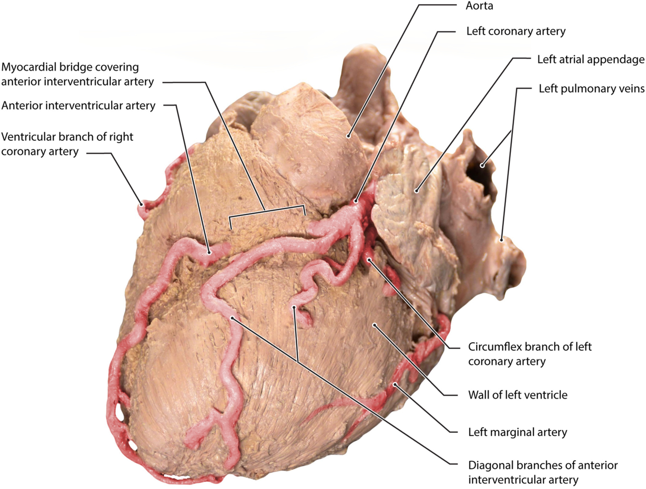
Her clinical examination showed mid-diastolic murmur at the apex. Her echocardiogram showed severe mitral restenosis with mitral valve area of 1.0 cm2 with calcified commissures and severely thickened leaflets. Pre-operative coronary angiogram showed intramyocardial left anterior descending (LAD) artery with myocardial bridge causing systolic and diastolic compression. Other coronaries were normal.
Surgical Procedure
She underwent mitral valve replacement with 27 mm bi-leaflet Carbomedics mechanical valve. She also had supracoronary artery myotomy, unroofing and marsupialization of the incised myocardium over LAD. Once the LAD was localized, by identifying the artery near the apex, the proximal LAD was exposed by incising the muscle and fat together with Pott’s scissors, keeping the lower scissor blade exactly on the anterior surface of the artery to minimize the chance of injuring the diagonals and entering into the right ventricular cavity. Each edge, including the full thickness of muscle and fat, was sutured in a running fashion with 6-0 Prolene. The suturing was started from the distal end in a continuous running manner. After reaching the other end, similar suturing was done in an over and over fashion in the reverse direction. This was repeated on the other side also. She came off bypass with normal ECG and stable hemodynamics. She made an uneventful postoperative recovery and was discharged from our care on the 7th postoperative day.
Discussion
Myocardial bridges (MB) are rarely observed, but well known, pathology of the major epicardial coronary arteries which are embedded in the overlying myocardial tissue. They are associated with myocardial ischemia and infarction, cardiac arrhythmias and sudden death. This entity is the cause of myocardial infarction with normal coronaries in some patients. Surgical myotomy reverses local myocardial ischemia and causes an increase in coronary blood flow. Supra-arterial decompressive myotomy treats the physiologic abnormality and corrects the congenital anatomic defect. Many techniques have been described and each has advantages and disadvantages. The problems that can be encountered during exposure of the intramyocardial LAD are (1) injury to the diagonal branches and the LAD itself and (2) entering into the right ventricle cavity. We describe our unique technique, which tackles these dangers effectively. This technique can also be used in conventional CABG with CPB or for off-pump CABG procedures. In addition, it helps to control bleeding from the cut edges.
