Cardiac perfusion in sickle cell disease

cardiac-perfusion

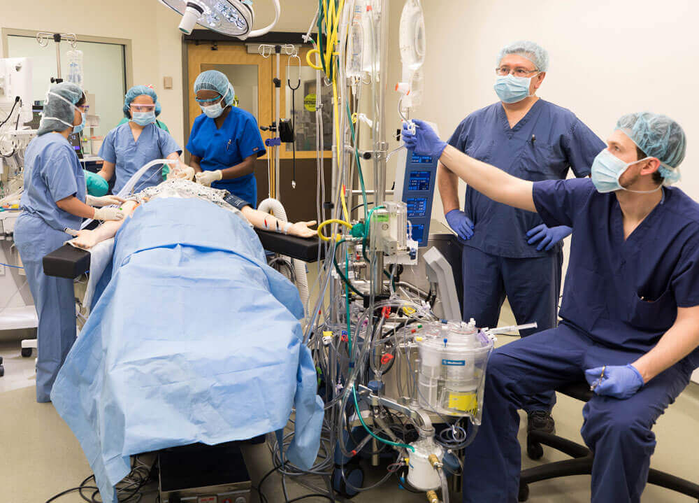
Cardiac operation in patients with sickle cell disease is a challenge to the surgical team. Performing cardiac surgery with the use of cardiopulmonary bypass (CPB) on patients with sickle cell disease requires extensive preoperative preparations and coordination from hematology, cardiac surgery, anesthetist, perfusion team and blood bank. CPB is commonly associated with hypothermia, hypoxia, hypoperfusion, and acidosis, which are pre-disposing factors that can trigger sickle cell crisis. Thus, careful attention to detail is vital to achieve the successful completion of the procedure safely, without triggering fatal vaso-occlusive episodes.

Here we describe our successful management of three sickle cell patients who underwent: 1) Aortic valve replacement with mitral valve repair 2) CABG 3) “Bentall” procedure in Acute Aortic dissection. Hemoglobin electrophoresis demonstrated HbS level of more the 40% in all these patients. Before surgery we decided to do perioperative exchange transfusion to

• promote perfusion,

• lower the incidence of perioperative sickling and

• lower the risk of perioperative multi-organ failure.

Hypothermia was prevented by maintaining the perfusate temperature at 35 degrees C. Hypoxia was avoided by maintaining hematocrit values above 24% and by keeping venous saturation above 60%. Frequent monitoring of ABG was done to maintain pH in the normal range. We did a partial exchange transfusion in the operating room for all three patients to reduce the HbS level to less than 20% without any perioperative transfusion. By using a separate reservoir and priming the oxygenator with heterologous blood we were able to reduce the intraoperative circulating HbS level to below 0% after exchange transfusion. This technique, combined with the use of continuous ultrafiltration on CPB, allowed the procedure to be performed without any complications. Cardiopulmonary bypass circuit was primed with 4 units of PRBC. The remaining prime consisted of 2 units of FFP, 100 ml of 20% albumin and sufficient Ringer lactate to complete the total circuit volume of 4 liter. The mixture was pre-oxygenated, warmed and buffered with sodium bicarbonate.

We performed exchange transfusion immediately before the initiation of CPB. After the patient received 3mg/Kg heparin, arterial and venous cannulation was performed. Four liter capacity reservoir was connected with a Y connector to the venous line of the CPB system to enable the perfusionist to collect the patient’s blood before CPB was initiated. Immediately before CPB was started about 3 liters of venous blood, which was estimated to be 2/3 of patient’s blood volume was allowed to rapidly flow in to the reservoir while patient was slowly transfused with 3 liters of solution from CPB system. Once 3 liters of blood was collected in the reservoir, venous flow was redirected to the oxygenator and normal CPB was initiated. Patient’s blood pressure was maintained at 60-70mmHg during exchange transfusion, with the use of vasoactive medications if required.

In the patient with aortic valve replacement and mitral valve repair, we used cold crystalloid cardioplegia. The theoretical advantage of crystalloid cardioplegia over blood cardioplegia is that it would avoid the possibility of sickling and vascular occlusion in coronary microvasculature. Hyperkalemia and hemodilution was avoided by sucking out the crystalloid cardioplegia from the right atrium. During CABG, the heart was initially arrested with 1200 ml of antegrade 4:1 blood–crystalloid cardioplegia maintained at 34 degrees C through the aortic root and in ‘Bentall’ procedure the same combination cardioplegia delivered through coronary ostia. Subsequent doses of cardioplegia at 34 degrees C were given retrograde through coronary sinus at 20-minute intervals. During CPB, flows were maintained at a minimum of 2.4L/m2. Venous saturation was kept above 60% and there was no acidosis during CPB. Arterial pressure was kept between 60 and 90mmHg.

Through pre-operative stabilization of the patient with respect to hemoglobin and use of initial exchange transfusion, customized cardioplegia and maintenance of blood gases, it was possible for CPB to be conducted safely in these sickle cell patients. The successful performance of these cases shows that planning, preparation and cooperation between team members and technical skill can overcome most operative challenges faced by sickle cell patients.
Chest wall tuberculosis

Cold abscess – a rare entity in the present era of anti-tubercular treatment

A 25-year-old student presented to the pulmonary medicine outpatient department with a low-grade fever, cough, and weight loss of 3kg in the preceding three months. He had no background atopy or usage of tobacco products. He had no shortness of breath, wheezing, skin, or joint disease. He denied a history of thyroid disease or high-risk sexual behavior. Enzyme-linked immunosorbent assay for HIV was negative. Sputum examination for acid-fast bacilli (AFB) sputum was positive for M. tuberculosis with no rifampicin resistance. He was started on standard four drugs anti-TB treatment with the symptomatic improvement of cough and weight gain.

He presented 2 months later with acute pain and swelling of the right axilla, of sudden onset. The swelling progressively increased over 7 days. On inspection, there was swelling of the right axilla. On palpation, the swelling was soft and fluctuant; there were no skin changes. Diagnosis of the tubercular cold abscess was made. Blood counts and liver functions were normal. Suspicion of irregular drug intake and drug-resistant TB was entertained. However, the patient denied irregular drug intake. He was referred to us for further management.

After stabilizing the patient, base line investigations were done. CT thorax showed multiple fluid collections with thick rim enhancing wall seen along the right posterolateral chest wall involving predominantly the right serratus anterior muscle and chest wall muscle. Largest pocket extended for about 12 cm in cranial caudal direction with width of 2.7cm. Superiorly fluid pocket/abscess pocket was seen to extend to the level of apex of lung. Anteromedially this abscess pocket was seen to extend along the extrapleural space on the medial aspect of the right 3rd rib. No obvious erosion/lytic area was noted in the surrounding bones.

Surgical Procedure

The patient was taken to operation room and under general anesthesia. The patient was put in right lateral position. Using a large spinal needle, aspiration was done and a sample of the abscess was taken for microbiological investigations. The abscess site was opened and drained. The abscess extended in the posterolateral aspect of the thoracic wall from axilla to lateral scapular border. The wound was closed over drains. He made an uneventful postoperative recovery and was discharged on the 6th postoperative day.

Discussion

The development of cold abscess is an important cause for poor clinical response or worsening in a patient on anti-tubercular treatment. It is seen in about 0.1% of musculoskeletal tuberculosis. Tubercular abscess of the chest wall is usually seen at the sternal margins or along the ribs. Mechanisms described include hematogenous dissemination, direct extension from lymphadenitis of the chest wall or suboptimal management of tubercular abscess of the chest wall and osteomyelitis of the ribs. Surgical excision of the affected tissue, under cover of anti-tubercular treatment, is believed to be the ideal strategy to prevent recurrence.

Triple valve surgery

A 31-year-old male, presented with history of dyspnea on exertion and chest pain (NYHA class III) and was diagnosed to have severe aortic stenosis and regurgitation, moderate to severe mitral stenosis and tricuspid valve stenosis. After initial stabilization and symptomatic improvement, the patient underwent coronary angiogram that showed normal coronaries. He was posted for triple valve surgery.

Surgical Procedure

Patient underwent triple valve procedure of aortic valve replacement with 21mm carbomedics mechanical valve, mitral valve replacement with 25mm carbomedics mechanical valve and tricuspid valve repair with 30mm MC3 ring. He made an uneventful postoperative recovery and was discharged after two weeks.

Discussion

Rheumatic heart disease (RHD) is a significant cause of cardiac operations in developing countries. Cardiac valve surgeries for RHD account for a significant portion of valve surgeries. Triple valve replacement (TVR) is deemed a complex and challenging choice for rheumatic heart disease (RHD) and carries significant mortality and morbidity. The challenge for surgeons is the prolonged cardio-pulmonary bypass (CPB) and myocardial ischaemic times. In-hospital mortality rate of about 10–12% has been reported. Advances in myocardial protection and CPB techniques, use of new generation valves, improvements in surgical techniques along with advances in perioperative and postoperative care can be credited for the improvement in early survival after triple valve surgery. Increased experience with triple valve procedures, advances in the treatment of postoperative heart failure, intensive patient follow-up, and extensive education on anticoagulation are also reasons for the improvement in patient survival.

Complete revascularization in coronary artery disease CABG x 9 grafts

A 61-year-old gentleman, known as hypertensive, diabetic and dyslipidemic, presented with chest pain and was diagnosed as Non ST Elevation Myocardial Infarction. His coronary angiogram revealed left main stenosis and chronic total occlusion of left circumflex and mid right coronary artery. His echocardiogram showed moderate left ventricular dysfunction and mild mitral regurgitation. He was taken up for urgent coronary artery bypass grafting.

Surgical Procedure

On cardiopulmonary bypass, he underwent CABG x 9 grafting. Left internal mammary artery anastomosed as a sequential graft to mid and distal left anterior descending artery. Additionally, he had 7 vein grafts anastomosed to two diagonals, two obtuse marginal and three grafts to posterolateral, posterior descending and ventricular branch of right coronary artery. Patient underwent an uneventful recovery postoperatively and was discharged on the seventh post-operative day.

Discussion

This case report highlights the advantages of the often maligned cardiopulmonary bypass in ensuring a complete revascularization in a patient with multiple severe diffuse triple vessel coronary artery disease. Revascularization of all the occluded coronary artery vessels, with as many as nine distal anastomosis, in diffuse coronary artery disease, is highly demanding. With high attrition rates of vein grafts, the utility of complete vascularization cannot be stressed enough. Off-pump revascularization is limited by quality of anastomosis and number of anastomosis.

Posterior mediastinal mass excision under standby cardiopulmonary bypass

A 40-year-old male presented with fever and cough of two weeks duration which did not settle with conservative management. CT chest revealed benign appearing large mediastinal non enhancing cyst of 7 x 8 x 9 cm causing pressure effect over the adjacent mediastinal vascular structures, esophagus, left bronchus and extending below and above aortic arch and occupying space between superior vena cava and ascending aorta.

Surgical Procedure

As a precaution, left femoral artery and vein was exposed and looped under local anesthesia. Airway was secured and lung isolation was achieved with double lumen tube under inhalation anesthesia. The patient underwent complete removal of posterior mediastinal tumor through left posterolateral thoracotomy in right lateral position. Complete excision of the mass was achieved without any damage to the surrounding structures. Lymph node masses were also removed. Patient had an uneventful postoperative recovery.

Conclusion

This case highlights some of the difficulties associated with the management of large mediastinal mass. Though rare, they cause compression of vital structures causing respiratory insufficiency or hemodynamic compromise. Detailed preoperative evaluation and planning is required to ensure a successful outcome. We were prepared to go on emergency cardiopulmonary bypass in case there was a loss of airway or hemodynamic collapse at induction. Additionally, postoperative evaluation of airway for bronchomalacia was done on spontaneous breathing prior to extubation.

Coronary artery disease in the young population

Noninvasive intervention / CABG / hybrid procedure

A 40-year-old expatriate presented with unstable angina. At the age of 31, he had stenting in LAD, obtuse marginal 1, obtuse marginal 2 and RCA. Repeat angiogram showed stent stenosis and progressive disease.

Surgical Procedure

He was taken up for coronary bypass surgery under cardiopulmonary bypass. He had CABG x 5 grafts. He had LIMA anastomosed distal to the stent in LAD and SVG anastomosed to first diagonal, OM1, OM2 and precrux RCA. There was no area in PDA or PLB to do the grafting. There were multiple stents in precrux RCA, PDA and posterolateral right. Long arteriotomy was done in RCA and all the stent with atheromatous material were pulled out distally and proximally. Coronary lumen was washed and grafting done. The patient had a smooth post-op recovery and was discharged on 7th post-op day.

Discussion

Diffuse coronary artery disease is a well-known fact in young Asian patients. Noninvasive coronary intervention is the treatment of choice in this group of patients. Total arterial grafting is not an easy task in diffuse distal disease. The cardiology team who treated him at the age of 35 did a good job. Later the disease progress and all the stents had progressive atheromatous lesions. Removal of a stent from the coronary is not without morbidity. In this patient, there was no alternative to find out a portion of a coronary artery for grafting. Hence, the stent was removed, cleaned the coronary and grafting done. It is advocated that surgery is possible even if all the coronaries are occupied with blocked stents.

Mitral valve repair – perfection

Mitral valve repair with CABG

A 55-year-old male with severe non-rheumatic mitral regurgitation, severe pulmonary hypertension, diabetes and coronary artery disease, was admitted for mitral valve repair and coronary bypass surgery. In view of his severe pulmonary artery hypertension, he was admitted and treated with Sildenafil (phosphodiesterase inhibitor) before taken up for surgery. His coronary angiogram showed critical occlusion in circumflex artery.

Surgical Procedure

Under cardiopulmonary bypass and cardioplegic arrest, mitral valve was inspected. He had prolapse of P2-P3 segments and chordae rupture at P2 segment. Quadrangular resection and repair of PML was done. Mitral annuloplasty was done using 30 mm Carpentier-Edwards Physio annuloplasty ring. SVG graft was done to OM2 circumflex artery. Came off bypass with stable rhythm and mild mitral regurgitation (MR) on TEE. De-cannulation done. While securing complete hemostasis, his blood pressure improved and then TEE showed moderate MR at P3 segment.

The question was whether to leave it and accept this MR or go back and get a perfect repair. The team decided to re-do the repair. Hence the patient was re-heparinized, cannulated, went on CPB, cardioplegia was given, left atrium was opened. P3 segment was found to be further prolapsed. This segment was re-fixed with neo-chordae and additional Alfieri stitch. Came off bypass. Post-op TEE showed no MR.

The patient became COVID positive in the post-op period and had an extended stay for further 10 days. Post-op follow-up showed no MR and the patient is now back at work.

Discussion

Degenerative mitral valve disease is the most common organic mitral valve pathology. Usually degenerative mitral valve pathology does not progress to clinically significant. But patients who develop symptoms attributable to MR have adverse prognosis and they need timely surgery. With surgery, essentially unavoidable in these patients, mitral valve repair has demonstrated superior short and long-term outcomes. We should try to avoid mitral valve replacement in these patients. Mitral valve repair has been consistently associated with lower risk of thromboembolism and improved survival. The method of mitral valve repair was evolved and improved. On the basis of operative findings, we resect and repair, consider neochordae insertion, placing an Alfieri stich, or doing a combination of these procedures. Once repair is attempted, we must almost always try to reach 100% perfection. At any cost, these patients should not have residual MR as this may progress and the patient may need re-do surgery.

In this case, we partially agreed to accept the mild MR in post-repair period, but within no time, TEE showed worsening MR. There should not be any hesitation to put the patient back on CPB and re-repair the mitral valve to get best results.

Congenital heart disease in adults with patent ductus arteriosus (PDA)

A 23-year-old female, was admitted to the hospital, with progressive shortness of breath on exertion, chest pain, palpitation, and fatigability over the last four years. Six months back she had undergone cesarean section (LSCS). During pregnancy, she had severe shortness of breath and it worsened as the pregnancy progressed. During LSCS she collapsed and had a cardiac arrest which was successfully resuscitated. Cardiology evaluation showed large PDA.

On clinical examination, she had continuous murmur over the upper part of the left chest. Her chest x-ray showed bilateral prominent bronchovascular marking. CT angiogram revealed large PDA with shunt. It also showed dilated left atrium and left ventricle with severe pulmonary congestion. Echocardiogram showed PDA with dilated left ventricle and LVEF 60%. Anatomically this PDA was not suitable for device closure.

Surgical Procedure

Through posterolateral thoracotomy, PDA was dissected and doubly ligated under hypotensive anesthesia. Additionally, she had a medium size vascular clip placed at the aortic side of the PDA. She had an uneventful recovery and was discharged on seventh post-operative day.

Discussion

Patent ductus arteriosus in the adult is an extremely rare phenomenon. This condition should have been diagnosed much earlier. Aneurysm, endocarditis and calcification are few of the serious sequelae as the age advances. There are various methods to treat this condition. Open surgical division or ligation has traditionally been an extremely successful and safe method of PDA closure. Comparative results are now achievable using transcatheter devices in majority of the cases.

Diffuse coronary artery disease options

Multiple coronary endarterectomy in diffuse complex coronary artery disease

A 53-year-old male, known patient of hypertension, hyperlipidemia and type I diabetes mellitus, presented to the hospital with acute-onset chest pain. After initial evaluation, he was diagnosed to have non-ST elevation myocardial infarction. Coronary angiogram showed severe triple vessel disease with chronic total occlusion of left anterior descending artery and significant diffuse lesions in the left circumflex artery and right coronary . He was referred for coronary artery bypass grafting.

Surgical Procedure

After initial evaluation, the patient underwent coronary artery bypass grafting on cardiopulmonary bypass. Diffuse disease of the left anterior descending artery (LAD) with lumen of less than 1mm necessitated long closed manual endarterectomy of proximal, mid and distal LAD. Left internal mammary artery was anastomosed to the distal LAD. Diffuse calcific disease with very narrow lumen of obtuse marginal branch of left circumflex was found. This required another closed manual endarterectomy of the same vessel followed by vein graft to it. Additional three vein grafts were placed to the posterior descending artery, posterolateral branch and diagonal branch after endarterectomy of those vessels. Electively this patient was put on intra-aortic balloon pump for 24 hours. Patient had an uneventful post-operative recovery with no ischemic events or hemodynamic instability. Post-operatively patient was discharged on the 6th post-operative day on antiplatelet and anti-coagulants in addition to routine postoperative medications.

Discussion

Diffuse coronary artery disease (CAD) as such cannot be tackled with percutaneous coronary intervention and is more likely to be referred for CABG. However, up to 25% of patients with diffuse CAD cannot be safely and successfully treated by standard CABG. Therefore, several techniques including coronary endarterectomy, which involves the removal of the atherosclerotic core from the coronary artery lumen through an arteriotomy, have evolved over the years. Despite initial adverse results (high operative mortality and perioperative myocardial infarction), several recent publications have shown that it can be safely performed with improvement in surgical technique and immediate post-operative anti-thrombotic measures. Coronary endarterectomy can assure complete revascularization preventing residual ischemia. We have described a patient in whom we did multiple endarterectomies successfully.

Cardiac surgery in COVID-19 pandemic

Right atrial mass in a post-COVID-19 patient

A 38-year-old lady presented to the hospital with shortness of breath and right sided chest pain. Symptoms were gradually progressive over few weeks. Her past history was significant with history of COVID-19 eight weeks prior. There were no remarkable clinical findings on examination.

Echocardiogram showed a right atrial mass of size 2.5 cm x 0.5 cm which was abutting the tricuspid valve. Patient had no other significant findings except moderate pulmonary artery hypertension on the echocardiography. She had no evidence of deep vein thrombosis in her lower limbs or pelvic veins and chest CT scan showed minimal post-COVID residual fibrotic bands. Her hematology work-up didn’t show any thrombotic predisposition.

She was started on anticoagulation with heparin initially and then warfarin. After four weeks of anticoagulation, there was no decrease in mass size and no significant improvement in her symptoms. It was decided to proceed with surgical excision in view of the above, suspicion of myxoma or organized thrombus and its risk of embolization.

Surgical Procedure

She underwent right atrial mass excision under cardiopulmonary bypass. Intraoperative transesophageal echocardiography confirmed the preoperative echocardiographic findings. Special attention had to be paid to careful cannulation of IVC without dislodging the mass as its attachment was close to the IVC-RA junction. The mass measured 2.0 x 2.0 cm. Part of the interatrial septum, that was excised with the mass, was repaired with pericardial patch.

Discussion

Multiple cases of right atrial mass following COVID-19 have been reported. An increased incidence of thrombotic events has been reported in patients with COVID-19 infection and pulmonary thromboembolism is one of the most important causes of clinical deterioration. We found a large thrombus in the right atrium after pneumonia caused by COVID-19, in a symptomatic patient in sinus rhythm with normal biventricular function. Prior reports have suggested improvement or prevention of deterioration in such patients with anticoagulation alone. We decided to proceed with surgical excision as the patient did not improve with four weeks of anti-coagulation.