Established leader in large, growing and resilient addressable market

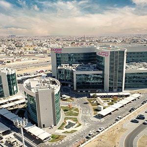
  • The largest private healthcare network in the UAE by bed count (1,660 beds) and private inpatient market share (19% in 2022)
  • The UAE, Oman & Saudi Arabia healthcare market represents a USD 69bn opportunity as of 2021, which is set to increase by a further 1.9x by 2031

Well-invested multi-brand network covering the full socio-economic spectrum

  • A portfolio of four well-known brands targeting the entire socio-economic spectrum, and creating a favourable negotiating position with insurance payers
  • Well invested network (AED 4.3bn total capex from inception) spanning 67 medical assets, including hospitals, medical centers and pharmacies
  • Strong investment in manpower has seen our team of clinically skilled and board-certified doctors grow by 53% since 2019 to 1,444 doctors

World-class super-specialty care offering driving patient yields

  • First mover and leading center in UAE for complex care, with transplant medicine, complex surgery, advanced oncology and other complex segments driving its patient yields growth (+9% ARR CAGR 2019-22)
  • Investing in healthcare partnerships to expand our complex service offering in the UAE in conjunction with internationally renowned providers

Accelerating digitization to drive operational and medical excellence

  • Digital health and digital transformation are key areas of investment across our network to optimize patient experience and maximize operational efficiencies
  • Our recently launched Patient Digital Channel continues to gain traction and now has  13% of total appointments

Expanding geographically through high-return and low-capex opportunities

  • Entry to high potential KSA market by introducing several low-capex, high-demand and locally-tailored healthcare products
  • Solid mid-term pipeline of completely de-risked and zero-capex public partnerships across MENA provides a huge bottom-line upside
  • Continued expanding our UAE healthcare infrastructure through adding 80 new in-demand specialized inpatient beds and five new medical centers in 2023

Cash-generative business model committed to delivering consistent shareholder return

  • Track record of strong financial performance and consistent growth, driven by the ramp-up of newly launched assets and supported by a focus on treating a higher-yielding patient mix in addition to cost optimization
  • Revenue CAGR of 17% over the past four years (FY 19-22), with impressive EBITDA CAGR (FY 19-22) of 28% and EBITDA margin expansion to 22% in FY 22
  • Robust margin expansion drivers coupled with compelling asset economics and strict capital discipline resulted in strong free cash flow generation capabilities (50% for H1 23)

Cardiac perfusion in sickle cell disease

cardiac-perfusion

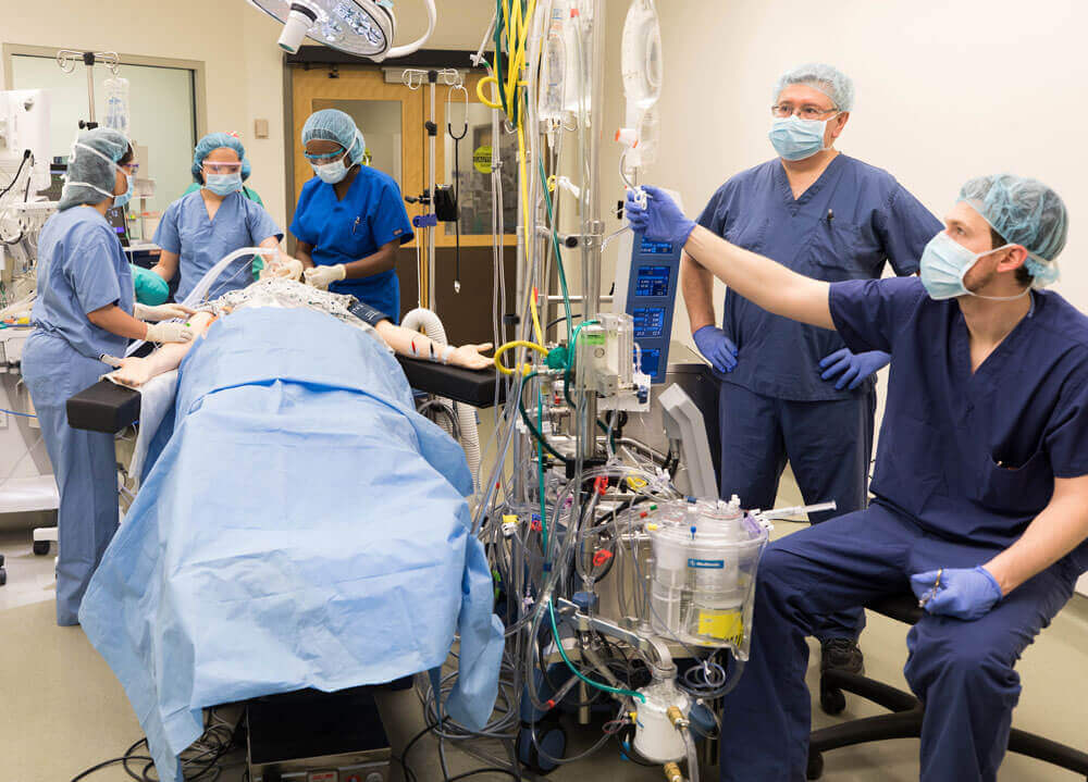
Cardiac operation in patients with sickle cell disease is a challenge to the surgical team. Performing cardiac surgery with the use of cardiopulmonary bypass (CPB) on patients with sickle cell disease requires extensive preoperative preparations and coordination from hematology, cardiac surgery, anesthetist, perfusion team and blood bank. CPB is commonly associated with hypothermia, hypoxia, hypoperfusion, and acidosis, which are pre-disposing factors that can trigger sickle cell crisis. Thus, careful attention to detail is vital to achieve the successful completion of the procedure safely, without triggering fatal vaso-occlusive episodes.

Here we describe our successful management of three sickle cell patients who underwent: 1) Aortic valve replacement with mitral valve repair 2) CABG 3) “Bentall” procedure in Acute Aortic dissection. Hemoglobin electrophoresis demonstrated HbS level of more the 40% in all these patients. Before surgery we decided to do perioperative exchange transfusion to

• promote perfusion,

• lower the incidence of perioperative sickling and

• lower the risk of perioperative multi-organ failure.

Hypothermia was prevented by maintaining the perfusate temperature at 35 degrees C. Hypoxia was avoided by maintaining hematocrit values above 24% and by keeping venous saturation above 60%. Frequent monitoring of ABG was done to maintain pH in the normal range. We did a partial exchange transfusion in the operating room for all three patients to reduce the HbS level to less than 20% without any perioperative transfusion. By using a separate reservoir and priming the oxygenator with heterologous blood we were able to reduce the intraoperative circulating HbS level to below 0% after exchange transfusion. This technique, combined with the use of continuous ultrafiltration on CPB, allowed the procedure to be performed without any complications. Cardiopulmonary bypass circuit was primed with 4 units of PRBC. The remaining prime consisted of 2 units of FFP, 100 ml of 20% albumin and sufficient Ringer lactate to complete the total circuit volume of 4 liter. The mixture was pre-oxygenated, warmed and buffered with sodium bicarbonate.

We performed exchange transfusion immediately before the initiation of CPB. After the patient received 3mg/Kg heparin, arterial and venous cannulation was performed. Four liter capacity reservoir was connected with a Y connector to the venous line of the CPB system to enable the perfusionist to collect the patient’s blood before CPB was initiated. Immediately before CPB was started about 3 liters of venous blood, which was estimated to be 2/3 of patient’s blood volume was allowed to rapidly flow in to the reservoir while patient was slowly transfused with 3 liters of solution from CPB system. Once 3 liters of blood was collected in the reservoir, venous flow was redirected to the oxygenator and normal CPB was initiated. Patient’s blood pressure was maintained at 60-70mmHg during exchange transfusion, with the use of vasoactive medications if required.

In the patient with aortic valve replacement and mitral valve repair, we used cold crystalloid cardioplegia. The theoretical advantage of crystalloid cardioplegia over blood cardioplegia is that it would avoid the possibility of sickling and vascular occlusion in coronary microvasculature. Hyperkalemia and hemodilution was avoided by sucking out the crystalloid cardioplegia from the right atrium. During CABG, the heart was initially arrested with 1200 ml of antegrade 4:1 blood–crystalloid cardioplegia maintained at 34 degrees C through the aortic root and in ‘Bentall’ procedure the same combination cardioplegia delivered through coronary ostia. Subsequent doses of cardioplegia at 34 degrees C were given retrograde through coronary sinus at 20-minute intervals. During CPB, flows were maintained at a minimum of 2.4L/m2. Venous saturation was kept above 60% and there was no acidosis during CPB. Arterial pressure was kept between 60 and 90mmHg.

Through pre-operative stabilization of the patient with respect to hemoglobin and use of initial exchange transfusion, customized cardioplegia and maintenance of blood gases, it was possible for CPB to be conducted safely in these sickle cell patients. The successful performance of these cases shows that planning, preparation and cooperation between team members and technical skill can overcome most operative challenges faced by sickle cell patients.
Chest wall tuberculosis

Cold abscess – a rare entity in the present era of anti-tubercular treatment

A 25-year-old student presented to the pulmonary medicine outpatient department with a low-grade fever, cough, and weight loss of 3kg in the preceding three months. He had no background atopy or usage of tobacco products. He had no shortness of breath, wheezing, skin, or joint disease. He denied a history of thyroid disease or high-risk sexual behavior. Enzyme-linked immunosorbent assay for HIV was negative. Sputum examination for acid-fast bacilli (AFB) sputum was positive for M. tuberculosis with no rifampicin resistance. He was started on standard four drugs anti-TB treatment with the symptomatic improvement of cough and weight gain.

He presented 2 months later with acute pain and swelling of the right axilla, of sudden onset. The swelling progressively increased over 7 days. On inspection, there was swelling of the right axilla. On palpation, the swelling was soft and fluctuant; there were no skin changes. Diagnosis of the tubercular cold abscess was made. Blood counts and liver functions were normal. Suspicion of irregular drug intake and drug-resistant TB was entertained. However, the patient denied irregular drug intake. He was referred to us for further management.

After stabilizing the patient, base line investigations were done. CT thorax showed multiple fluid collections with thick rim enhancing wall seen along the right posterolateral chest wall involving predominantly the right serratus anterior muscle and chest wall muscle. Largest pocket extended for about 12 cm in cranial caudal direction with width of 2.7cm. Superiorly fluid pocket/abscess pocket was seen to extend to the level of apex of lung. Anteromedially this abscess pocket was seen to extend along the extrapleural space on the medial aspect of the right 3rd rib. No obvious erosion/lytic area was noted in the surrounding bones.

Surgical Procedure

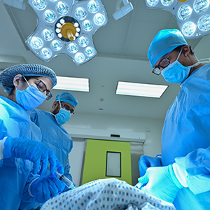
The patient was taken to operation room and under general anesthesia. The patient was put in right lateral position. Using a large spinal needle, aspiration was done and a sample of the abscess was taken for microbiological investigations. The abscess site was opened and drained. The abscess extended in the posterolateral aspect of the thoracic wall from axilla to lateral scapular border. The wound was closed over drains. He made an uneventful postoperative recovery and was discharged on the 6th postoperative day.

Discussion

The development of cold abscess is an important cause for poor clinical response or worsening in a patient on anti-tubercular treatment. It is seen in about 0.1% of musculoskeletal tuberculosis. Tubercular abscess of the chest wall is usually seen at the sternal margins or along the ribs. Mechanisms described include hematogenous dissemination, direct extension from lymphadenitis of the chest wall or suboptimal management of tubercular abscess of the chest wall and osteomyelitis of the ribs. Surgical excision of the affected tissue, under cover of anti-tubercular treatment, is believed to be the ideal strategy to prevent recurrence.

Triple valve surgery

A 31-year-old male, presented with history of dyspnea on exertion and chest pain (NYHA class III) and was diagnosed to have severe aortic stenosis and regurgitation, moderate to severe mitral stenosis and tricuspid valve stenosis. After initial stabilization and symptomatic improvement, the patient underwent coronary angiogram that showed normal coronaries. He was posted for triple valve surgery.

Surgical Procedure

Patient underwent triple valve procedure of aortic valve replacement with 21mm carbomedics mechanical valve, mitral valve replacement with 25mm carbomedics mechanical valve and tricuspid valve repair with 30mm MC3 ring. He made an uneventful postoperative recovery and was discharged after two weeks.

Discussion

Rheumatic heart disease (RHD) is a significant cause of cardiac operations in developing countries. Cardiac valve surgeries for RHD account for a significant portion of valve surgeries. Triple valve replacement (TVR) is deemed a complex and challenging choice for rheumatic heart disease (RHD) and carries significant mortality and morbidity. The challenge for surgeons is the prolonged cardio-pulmonary bypass (CPB) and myocardial ischaemic times. In-hospital mortality rate of about 10–12% has been reported. Advances in myocardial protection and CPB techniques, use of new generation valves, improvements in surgical techniques along with advances in perioperative and postoperative care can be credited for the improvement in early survival after triple valve surgery. Increased experience with triple valve procedures, advances in the treatment of postoperative heart failure, intensive patient follow-up, and extensive education on anticoagulation are also reasons for the improvement in patient survival.