Giant left and right atrium

Surgical correction in re-do mitral valve surgery

Rheumatic heart disease (RHD) is the only preventable cardiovascular disease which causes significant morbidity and mortality particularly in low- and middle-income countries. The need for surgery for severe RHD is clear with a significant lack of access to surgical and percutaneous intervention in regions of the greatest global need. Surgery for RHD should be timeous to result in the best possible outcome. In advanced RHD, heart failure, atrial fibrillation and endocarditis can add to the complexity of the surgery.

History

A 45-year-old Sudanese lady, with history of mitral valve repair done 20 years prior, presented with dyspnea on exertion, chest pain and severe pulmonary artery hypertension. Echocardiogram showed severe mitral stenosis, moderate mitral regurgitation, moderate tricuspid regurgitation, severe pulmonary artery hypertension, enlarged right atrium and giant left atrium. Coronary angiogram showed normal coronaries.

Surgical Procedure

Under cardiopulmonary bypass and cardioplegic arrest, she underwent redo mitral valve replacement (29mm carbomedics standard mechanical valve), tricuspid valve repair with Annuloplasty (32mm Edwards MC3 Annuloplas-ty ring), left atrial appendage closure from left atrium, left atrial size reduction with plication, and right atrial size reduction. Came off CPB in the first attempt, in sinus rhythm. Patient made a gradual uneventful recovery post-op and was discharged home on the tenth post-operative day.

Discussion

RHD remains a neglected cardiovascular disease, which causes significant morbidity and mortality in low- and middle-income countries. Timing of surgical intervention is crucial for a favorable outcome. Our patient with history of previous mitral valve repair, presented relatively late with compounding risk factors of cardiac failure, atrial fibrillation, pulmonary artery hypertension, tricuspid regurgitation and giant left atrium, in addition to the primary pathology of mitral valve disease. Giant left atrium (GLA) is a condition defined when the left atrial diameter exceeds 65 mm. It is commonly associated with mitral valve regurgitation due to excess intracavitary pressure resulting in strain and dilation of the left atrial chamber. The enlarged left atrium leads to expansion of left atrial volume, which in turn can place pressure on the main bronchus, lung and left ventricle with corresponding cardiopulmonary embarrassment. Because GLA can increase the risk of sudden death, its existence merits surgical intervention. Partial resection of inferior and or superior left atrial wall, is the most common surgical technique. With the evolution of atrial fibrillation surgery, atrial size matters and is determinant of long term performance following successful ablation. Surgical management of GLA achieves good clinical outcome with respect to cardiopulmonary performance including restoration of sinus rhythm among patients suffering from atrial fibrillation.

In patients who undergo mitral valve surgery with tricuspid regurgitation or tricuspid annular dilation, performing concomitant tricuspid valve repair lowered risk for progression of tricuspid regurgitation. There is broad agreement that when a patient has severe tricuspid regurgitation, then that valve should be repaired. However, there is significant uncertainty in how to manage moderate or less tricuspid regurgitation. There are some data from observational studies that failure to manage less than severe tricuspid regurgitation is associated with decreased survival and heart failure. It is a common debate among surgical team members as to what to do with leaking tricuspid valve during mitral valve surgery. It is recommended that the tricuspid valve annulus be intervened based on function (i.e., degree of tricuspid valve regurgitation), diameter size, significant right ventricular dilation or dysfunction. This patient presented all the difficulties associated with rheumatic valve surgery including cardiac failure, atrial fibrillation, associated valve lesion, pulmonary artery hypertension and giant left atrium. Careful assessment, pre-operative optimization, meticulous surgical technique and optimal post-operative management played an important role in successful outcome.

Bentall procedure and mitral valve repair

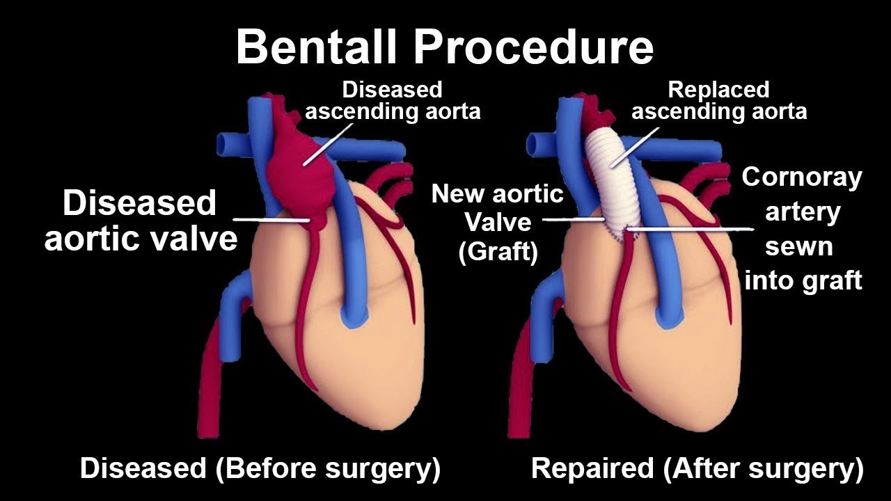
A 34-year-old male from Cameroon, with Marfanoid features was referred for aortic root replacement and mitral valve surgery. His echocardiogram showed severe ascending aortic dilatation more than 8 cm and severe mitral regurgitation. Preoperative TEE showed dilated aortic annulus and ascending aorta, dilated mitral annulus, with severe AR and MR. He was planned for Bentall procedure and mitral valve repair.2

Surgical technique

Under cardiopulmonary bypass and cardioplegic arrest, ascending aorta and the aortic valve was replaced with 25 mm Carbomedics valve bearing conduit with coronary implantation. Mitral valve showed normal leaflets with dilated annulus. Mitral annuloplasty was done using a 32 mm Carpentier-Edwards Physio ring. Post-op TEE showed no MR and normally functioning aortic prosthesis. He had a smooth post-op recovery and was discharged on seventh post-op day.

Discussion

Co-existing aortic root and mitral valve pathology is increasingly recognized among patients undergoing cardiac surgery. This combination of pathologies is particularly common in patients with connective tissue disorders such as Marfan syndrome. Aortic root replacement with mitral valve repair or replacement are the surgical options to treat these patients. However, the combination of the mitral valve operation and an aortic root procedure is more demanding than either major operation alone and knowledge regarding the clinical outcome is limited. The most common pathologies are root aneurysm without dissection and degenerative mitral valve disease. Combined aortic root and mitral valve operations are safe and associated with excellent long-term results.

Myocardial bridges – when to operate

myocardio

A 43-year-old lady, with history of childhood rheumatic fever, presented with history of gradually progressive dyspnea on exertion, palpitation and recent onset of chest pain. She had undergone percutaneous mitral balloon valvotomy fifteen years back for mitral stenosis. She had no other significant history.

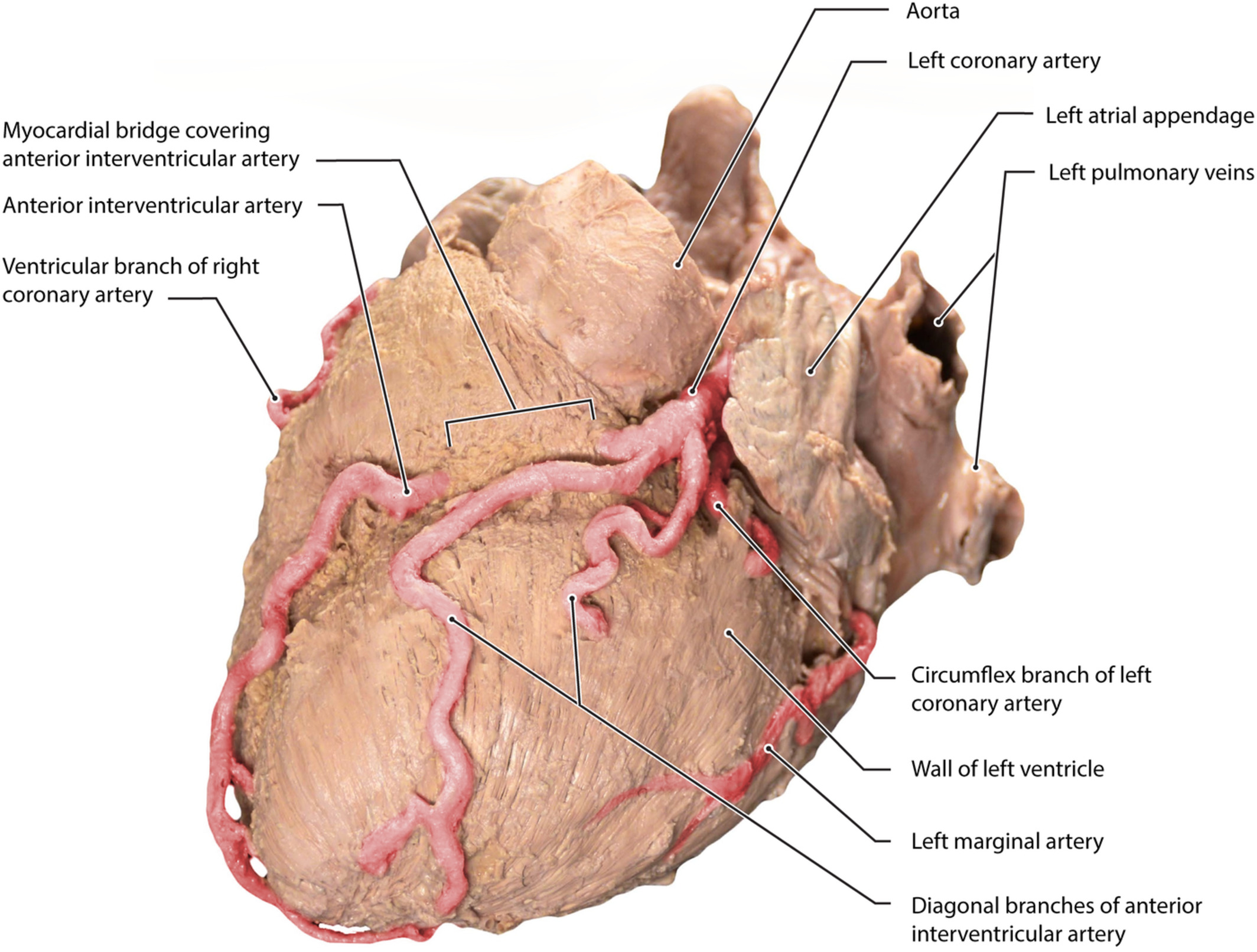
Her clinical examination showed mid-diastolic murmur at the apex. Her echocardiogram showed severe mitral restenosis with mitral valve area of 1.0 cm2 with calcified commissures and severely thickened leaflets. Pre-operative coronary angiogram showed intramyocardial left anterior descending (LAD) artery with myocardial bridge causing systolic and diastolic compression. Other coronaries were normal.

Surgical Procedure

She underwent mitral valve replacement with 27 mm bi-leaflet Carbomedics mechanical valve. She also had supracoronary artery myotomy, unroofing and marsupialization of the incised myocardium over LAD. Once the LAD was localized, by identifying the artery near the apex, the proximal LAD was exposed by incising the muscle and fat together with Pott’s scissors, keeping the lower scissor blade exactly on the anterior surface of the artery to minimize the chance of injuring the diagonals and entering into the right ventricular cavity. Each edge, including the full thickness of muscle and fat, was sutured in a running fashion with 6-0 Prolene. The suturing was started from the distal end in a continuous running manner. After reaching the other end, similar suturing was done in an over and over fashion in the reverse direction. This was repeated on the other side also. She came off bypass with normal ECG and stable hemodynamics. She made an uneventful postoperative recovery and was discharged from our care on the 7th postoperative day.

Discussion

Myocardial bridges (MB) are rarely observed, but well known, pathology of the major epicardial coronary arteries which are embedded in the overlying myocardial tissue. They are associated with myocardial ischemia and infarction, cardiac arrhythmias and sudden death. This entity is the cause of myocardial infarction with normal coronaries in some patients. Surgical myotomy reverses local myocardial ischemia and causes an increase in coronary blood flow. Supra-arterial decompressive myotomy treats the physiologic abnormality and corrects the congenital anatomic defect. Many techniques have been described and each has advantages and disadvantages. The problems that can be encountered during exposure of the intramyocardial LAD are (1) injury to the diagonal branches and the LAD itself and (2) entering into the right ventricle cavity. We describe our unique technique, which tackles these dangers effectively. This technique can also be used in conventional CABG with CPB or for off-pump CABG procedures. In addition, it helps to control bleeding from the cut edges.

Ventricular septal rupture

A 54-year-old male came with history of chest pain of 3 weeks duration. He was a known diabetic and hypertensive. He was not on any treatment at that time. His symptoms worsened after three weeks, when he presented to the hospital. On evaluation, he was detected to have ventricular septal rupture following acute coronary syndrome. He had features of pulmonary edema and congestive heart failure. Echocardiogram showed moderate LV dysfunction with ejection fraction of 35%, mild MR and 18mm ventricular septal rupture at the apex with dyskinetic apex. Coronary angiogram showed severe triple vessel dis-ease. After initial stabilization, he was taken up for surgery after a week when his renal function started deteriorating.

Surgical Procedure

He underwent ventricular septal repair using Gore-Tex patch and coronary artery bypass grafting (CABG x5) under cardiopulmonary bypass and intra-aortic balloon pump counter-pulsation. His post-operative period was stormy with repeat episodes of cardiac failure requiring prolonged inotropic support and ICU care, which was further complicated by renal dysfunction that was managed conservatively.

Discussion

Ventricular septal rupture repair is a rare surgical intervention with a high complication and mortality rate. On the other hand, if it is left untreated, it has a high mortality. In the acute phase, because the remaining septum is mushy necrotic muscle, they are not receptive to sutures. The hospital survival after ventricular septal rupture repair varies between 30–47%. These high mortalities have to be set against the mortality without surgery which is 94%.

Surgical principles of VSR closure include hypothermic cardiopulmonary bypass with myocardial protection, trans-infarction approach to the VSR, trimming of infarcted muscle around the VSR, closure of the VSR with a patch to avoid tension and closure of the ventricle without tension, with Teflon felt using buttressed sutures. Closure of ventricular septal rupture following myocardial infarction is an infrequent operation with a very high operative risk. Nonetheless, early surgical intervention offers the only realistic chance of survival and this opportunity should not be denied to patients. Immediate intra-aortic balloon counterpulsation provides some haemodynamic optimization while preparations are made for surgery.

The good long term outcome for survivors makes the high early mortality worthwhile. Transcatheter closure of ventricular septal rupture in the acute setting has been reported. This may provide temporary hemodynamic relief and therefore allow surgical closure after the infarcted myocardium around the rupture has had time to fibrose. With the current devices, closure can be very difficult

Discussion

Ventricular septal rupture repair is a rare surgical intervention with a high complication and mortality rate.

In the acute phase, because the remaining septum is mushy necrotic muscle, they are not receptive to sutures. The hospital survival after ventricular septal rupture repair varies between 30–47%. These high mortalities have to be set against the morality without surgery, which is 94%. Surgical principles of VSR closure include hypothermic cardiopulmonary bypass with myocardial protection, trans-infarction approach to the VSR, trimming of infarcted muscle around the VSR, closure of the VSR with a patch to avoid tension and closure of the ventricle without tension, with Teflon felt using buttressed sutures.

Closure of ventricular septal rupture following myocardial infarction is an infrequent operation with a very high operative risk. Nonetheless, early surgical intervention offers the only realistic chance of survival and this opportunity should not be denied to patients. Immediate intra-aortic balloon counterpulsation provides some haemodynamic optimisation while preparations are made for surgery. Another adjunct for those patients who are severely hemodynamically compromised are ventricular assist devices. As the technology of devices advances they should become increasingly available and increasingly adaptable; one can imagine the situation where a temporary device will maintain a patient’s haemodynamics while the edges of the septal rupture fibrose sufficiently to allow a definitive surgical closure later.

Coronary artery perforation during coronary angiogram – surgical challenges

An 82-year-old UAE national, who underwent cardiac catheterization procedure at Al Ain Hospital, developed coronary artery perforation during cardiac catheterization. Emergency covered stent grafting was done and perforation was closed. But unfortunately, the patient developed acute cardiac tamponade and needed resuscitation. Initially 100 ml of blood was drained from the pericardium percutaneously, but the patient’s condition did not improve and he went into cardiogenic shock. He was intubated and IABP was introduced with heavy inotropic support.

The patient was shifted for emergency cardiac surgery and drainage of cardiac tamponade. The patient’s history revealed ischemic heart disease, diabetes, hypertension, past history of smoking, with recent unstable angina. He was admitted with chest pain as NSTEMI at Al Ain Hospital. He had previous coronary angiogram in 2019 which showed double vessel disease and stenting was done to LAD and circumflex arteries. This time, in view of his unstable angina, he was taken up for diagnostic angiography and possible intervention. On admission to our hospital, he was in cardiogenic shock, on heavy inotropic support, ventilated and on IABP with poor hemodynamics. Preoperative transesophageal echocardiogram in the operation theatre revealed significant cardiac tamponade, ejection fraction 25%, dyskinetic apical and septal region, calcified aortic valve and presence of a large atheroma in distal aortic arch. He was taken up for emergency CABG and pericardial drainage was done. On opening the pericardium, almost 200 mL blood was drained and his hemodynamics improved. In view of the large mobile atheroma in the aortic arch, it was decided to do the coronary bypass grafting on OFF PUMP beating heart. On opening the LAD, there were fresh clots in both distal and proximal areas. A size 3 Fogarty balloon catheter was introduced to both proximal and distal LAD to remove the clots. Then single saphenous vein graft was done to mid LAD. He remained in on IABP and inotropic support with stable hemodynamics. All the supports were weaned off on the second post-operative day and IABP was removed. He was discharged on 12th post-operative day with no neurological deficit.

Discussion

Coronary artery perforation during cardiac interventions, is a rare complication in the present era. Advanced hardware, safety measures and highly experienced interventional cardiologists has brought down this complication to less than 0.3%. But when it occurs, if further interventional procedures fail, surgical treatment is the only alternative. For that, surgical team and the facility should be available at short notice to save the life of the patient.

Combined Bentall procedure with NUSS operation

Combined Bentall procedure with NUSS operation

A 45-year-old known patient of Marfan syndrome, presented with history of progressive shortness of breath, palpitations and easy fatigability over a period of 6 months.

Clinical Findings

  • Pectus excavatum (since childhood) 6.5 cm depth.
  • Early diastolic murmur over aortic area.
  • Marfan syndrome features present.

Primary Investigations & Findings

  • 2-D transthoracic echo: Severe aortic regurgitation and ascending aortic aneurysmal dilatation of 7 cm
  • CT angiogram: Fusiform
  • aneurysm of ascending aorta measuring 7 cm
  • CT Thorax: Ascending aortic aneurysm and sternal compression of right ventricle. Reduction of right lung volume, mediastinal shift to the left

Diagnoses

Ascending aortic aneurysm with severe aortic regurgitation and pectus excavatum.

Management Plan

Bentall procedure was done for the ascending aortic aneurysm and severe aortic regurgitation. Additional NUSS Bar fi¬xation was done for correction of pectus excavatum.

Aspects of Challenge in the Case

1) Sternotomy posed risk of RV tear due to its compression by the pectus excavatum.

2) Risk of aneurysm rupture during sternotomy.

3) Bentall procedure and its attendant unique risks such as bleeding and neurological sequela, etc.

4) NUSS Bar procedure for pectus excavatum correction, in the back ground of major cardiac surgery, as combined procedure.

5) Modified NUSS Bar fixation with sternal osteotomies.

Follow-up

Chest wall deformity corrected. He is on regular anti-coagulation and doing well.

Dextrocardia and coronary artery disease Off-pump CABG in dextrocardia and carotid endarterectomy

Dextrocardia with situs inversus is a rare congenital abnormality with an incidence of only 1:10,000. The incidence of coronary artery disease in this condition is similar to that in the general population.

History

A 66-year-old patient with a diagnosis of left main coronary artery disease and triple vessel disease, was referred for coronary bypass surgery. He was a known case of dextrocardia with situs inversus. He presented with unstable angina. He was a known case of stage 3 chronic renal disease, hypertension and diabetes mellitus. Carotid scan showed bilateral carotid artery stenosis with critical block in the left carotid artery. His coronary angiogram revealed triple vessel disease and dextrocardia.

Surgical Procedure

In preparation for coronary artery bypass surgery, he underwent left carotid endarterectomy. He had an uneventful recovery with no neurological deficit. After six weeks, he was taken up for CABG. Chest was opened through median sternotomy approach. Skeletonized right internal mammary artery (RIMA) was harvested and found to be good size and length and three lengths of long saphenous vein were harvested. Heart was stabilized with a tissue stabilizer device and RIMA was anastomosed to the LAD from the right side. Surgeon came on the left side and stabilized the heart for SVG to ramus intermedius, circumflex PDA and right acute marginal. All the distal anastomoses on those vessels were done from the left side and proximal anastomosis from the right side. Intra and post-op period was uneventful. His serum creatinine went up early post-op period and came down to his pre-op levels in five days’ time. Post-op echo showed normal LV function. He was discharged on 10th post-operative day.

Discussion

Performing CABG in dextrocardiac patient is more difficult and technically challenging as it is not routinely performed. There are few reported cases of CABG in dextrocardia and even lesser number had off-pump approach. Myocardial revascularization in dextrocardia can successfully be achieved with RIMA to the LAD and SVG to other vessels.

Mitral valve replacement and tricuspid valve repair in a patient with sickle cell disease

A 77-year-old American national was admitted with history of palpitation, chest pain and shortness of breath. He was very fragile and weighed only 48 kg. He was diagnosed to have severe rheumatic mitral regurgitation, severe tricuspid regurgitation, atrial fibrillation, and left ventricular failure. He had an attempted mitral clip procedure in another hospital, which failed. He was a known case of stage 4 chronic renal failure, sickle cell anemia, thrombocytopenia, diabetes mellitus and visual defects as tubular vision. The patient had multiple history of sickle cell crisis. Two years back he was admitted with septicemia, comatosed, ventilated and had tracheostomy in a hospital in the USA. This case was discussed in our heart failure clinic and accepted for the high risk procedure.

Surgical technique

All precautions were taken to deal with problems of sickle cell disease on cardiopulmonary bypass. The patient had exchanged transfusion before instituting cardiopulmonary bypass. He was hydrated well and 2.5 L of blood was removed at the beginning of CPB to a separate cardiotomy reservoir and discarded. CPB was started with blood prime to replace the lost volume and kept on normo-thermic. Operating room temperature was kept at 25 degrees C. Ultra-filtration was also used. The mitral valve was replaced with 27 mm bioprosthetic valve and tricuspid valve was repaired with 30 mm MC3 annuloplasty ring. Transatrial closure of the left atrial appendage was done using two-layer suture. The patient came off bypass with stable hemodynamics on minimal inotropic support. In the postop period, all precautions were taken to avoid sickle cell crisis. He had a smooth outcome and was discharged on 15th post-op day. He is being followed up in our clinic and is doing well.

Discussion

Patient with pre-op high morbidity underwent a very high risk procedure. This case was initially managed in another cardiac institute and decided not to subject him for open heart surgery due to his comorbidities. Hence mitral clip procedure was attempted, but failed. Following this, he had multiple admissions with pulmonary edema. The family support and the patient’s motivation was great and encouraged us to take him up for surgery. This helped to achieve a good result.

Cardiogenic shock

Acute rupture of anterior papillary muscle of mitral valve – emergency institution of cardiopulmonary bypass in the ICU

History

A 51-year-old male patient, known case of hypertension, hyperlipidemia, and diabetes mellitus type II (not on regular medication), presented to another hospital after collapse at work. He was having chest pain for previous 3 days, had not sought any help, till he collapsed. In the Emergency room he was having acute shortness of breath and hypoxia. He was intubated in the Emergency Room. He had a massive pulmonary edema. His investigations showed highly elevated cardiac markers (Tro-ponin T > 3000 pg/mL and Pro BNP > 3000 pg/mL). He was taken up for coronary angiogram, which showed triple vessel disease with acute thrombosis of RCA. Balloon Angioplasty was done to RCA. In view of his cardiogenic shock and severe hypo-tension, despite heavy inotropic support (Nor Adrenaline and Adrenaline), he was put on IABP. As his condition did not improve, cardiac surgery team at Burjeel was consulted and immediate transfer was advised. On arrival, he was in renal shut-down and was developing hepatic dysfunction. Acidosis persisted and despite corrective measures and adjusting ventilator parameters, his condition deteriorated. He was given multiple doses of Phe-nylephrine injection and a Methyl-ene Blue infusion was started. His heart rate remained above 140/min. Transthoracic echocardiogram showed fair right and left ventricular contractility. Despite very poor prognosis for any intervention, the relatives granted permission and requested to go ahead with any procedures. A bedside VA ECMO was instituted with centrifugal pump through femoral cannulation. He developed VT & VF, during the procedure, needing defibrillation. Gradually he got stabilized with ECMO, the heavy dose of inotropic support could be reduced with mean pressure of 60-65 mmHg. His heart rate came down to 70-80/min in normal sinus rhythm. TEE was done. Severe Mitral Regurgitation was observed with anterior papillary muscle rupture. His condition was explained to the relatives. In view of the refractory cardiogenic shock, unstable hemodynamics, despite IABP and ECMO, it was decided to proceed for Emergency CABG + MVR as a desperate life-saving measure.

Surgical Procedure

Intra Operatively ECMO was converted to Cardio Pulmonary Bypass. CABG x 3 and Mitral Valve was replaced with 25mm size CE bioprosthetic valve. As anticipated, he had difficulty in coming off cardiopulmonary bypass; hence, VA ECMO was reinstituted through the same femoral access, which was used for CPB.

He had significant bleeding for the next 24 hours on ECMO. He had persistent refractory hyperkalemia (serum potassium above 8 mmol/L). It did not respond to conventional measures to lower the potassium. CRRT was started in the ICU. Patient gradually worsened despite ECMO, IABP and heavy inotropes and expired on the 4th post-operative day.

Conclusion

1) Quick action is needed in saving patients with post infarct papillary muscle rupture and cardiogenic shock. In this patient, papillary muscle rupture and resultant acute severe MR could, unfortunately, be recognized only afer the patient was stabilized on ECMO, with his heart rate settling and with the help of TEE.

2) Multi-organ failure sets in very fast worsening the outcome and hence timely intervention is essential to salvage the myocardium and the patient.

3) The team should be prepared to take care of the very sick patient in the post-op period.

4) Family should be briefed about the situation and possible worst outcome.

In this patient, earlier ECMO might have helped to save the patient. In its absence at the referral unit, a mobile retrieval ECMO unit from our institution could have made it possible. Unfortunately such a mobile team was not existent at that time. Since then we have instituted the same.

Dor procedure for left ventricular reconstruction

Surgical ventricular restoration by means of Dor procedure is a surgical option in patients with coronary artery disease and post infarcular left ventricular aneurysm. This procedure gives a better outcome in those patients with poor left ventricular ejection fraction. Traditional cardiac surgical methods may have limited benefits in these types of patients. The Dor procedure excludes akinetic or dyskinetic portion of the anterior wall and septum, reshapes the LV with a stitch that encircles the transitional zone between normal contractile myocardium and aneurysmal scar tissue, and uses a patch to reestablish ventricular wall contour. By this technique, LV size and geometry is improved, reduces wall tension and enhances overall systolic function. Coronary bypass surgery is almost always performed with this procedure.

A 52-year-old American national was referred to cardiac surgery from outside the country with class IV symptoms. He was getting treatment for his hypertension and diabetes mellitus. His coronary angiogram showed left main stem stenosis and total occlusion of the LAD. Echocardiogram showed left ventricular aneurysm with LV clot, and no mitral regurgitation. His LV ejection fraction was 20%. He was scheduled for Dor procedure and CABG.

Surgical Procedure

Under cardiopulmonary bypass and cardioplegic arrest, the aneurysm was incised parallel to the LAD and clots were removed. A purse-string suture using 2-0 prolene was placed around the circumference of the scar at the transitional zone and tied down to determine size of the new ventricular opening. A Gore-tex patch was then sutured to the ventricular opening with interrupted 2-0 prolene suture. A second layer of continuous suture using 2-0 prolene is done. The edges of the ventricular free wall were then closed over the patch with a running 2-0 prolene supported with Teflon felt. The distal coronary anastomosis was done by saphenous vein to diagonal 1 and obtuse marginal 1. Left internal mammary was anastomosed to the proximal LAD. Proximal anastomosis was done to the ascending aorta with a side biting clamp. The patient was then weaned from CPB in a standard fashion. TEE was used to assess filling contractility and mitral valve function. He was electively put on IABP. Postoperative TEE showed improved LV contractility.

Discussion

The operative goal of the Dor procedure is to achieve complete coronary revascularization, reduce LV volume and restore its shape. The Dor procedure improves quality of life and survival in patients with left ventricular aneurysm. Also , complete revascularization is mandatory for a good outcome.